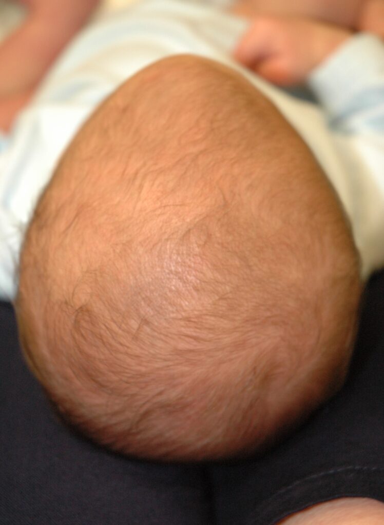
What is it?
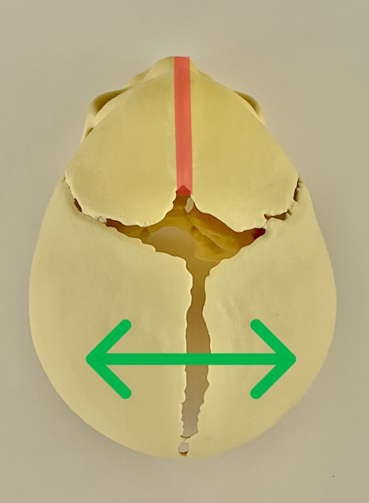
The metopic suture is a skull expansion joint running vertically down the middle of the forehead (shown in red in the image below). When it prematurely fuses, the forehead cannot widen, causing an abnormally narrow, pointed forehead shape.
What to look for?
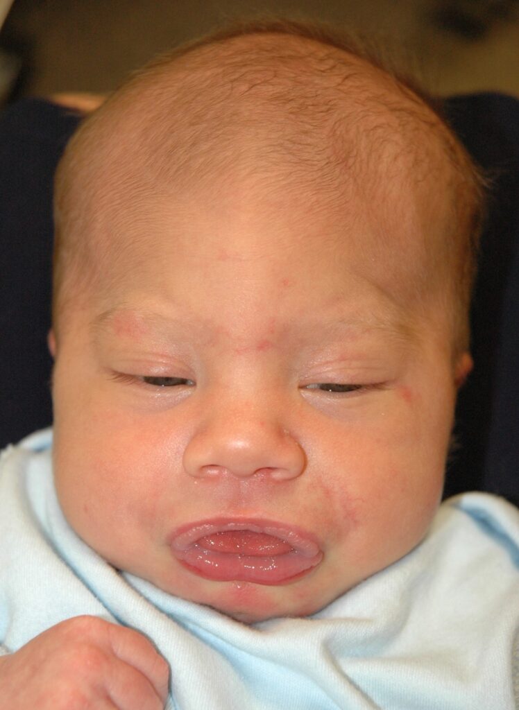
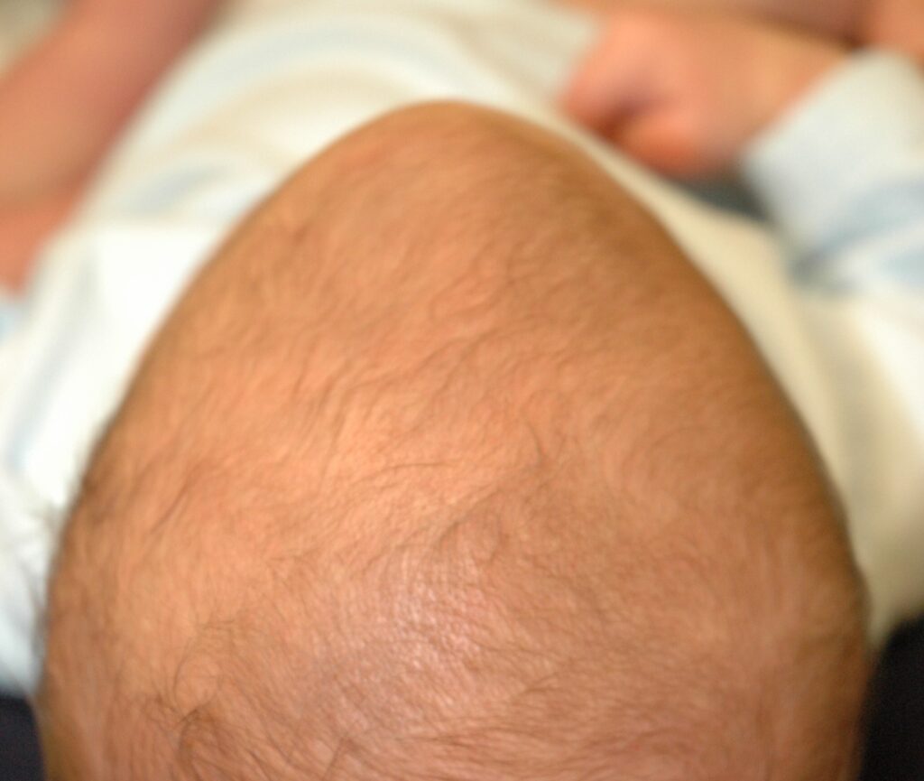
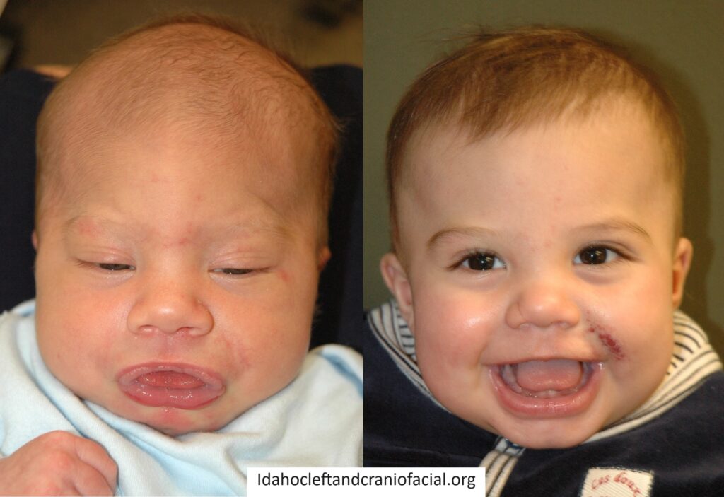
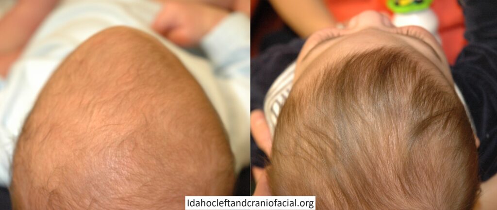
The forehead may have a vertical ridge and appear pointed when viewed from above. On overhead view, the skull often appears triangular, as the back of the skull flares wider. The forehead can also appear pinched and the eyes too close together.
Why is surgery recommended?
When the skull cannot grow as it should, it becomes progressively abnormal in appearance. Sometimes, intracranial pressure can become elevated. Surgery is recommended to normalize appearance, limit negative social interactions and reduce the risk of developmental concerns from elevated pressure.
Before and After Cranioplasty Surgery
Frequently Asked Questions
The skull sutures can be thought of as expansion joints. These expansion joints play a critical role in allowing the skull to grow quickly when the brain is growing rapidly in the first 12 to 18 months of life. When one of these expansion joints fuses, this restricts growth in the direction perpendicular to the suture. When the metopic suture, which runs down the middle of the forehead, is involved, the forehead cannot widen as it wants to. The forehead appears narrow and ridged, the temporal area appears pinched and the eyes can appear too close together. But the brain is resilient and will find a way to grow, usually pushing the posterior skull wider, giving the characteristic flared appearance of trigonocephaly, or triangular shaped skull.
There are 2 surgical approaches to treating metopic craniosynostosis – a minimally invasive approach and a traditional open surgical approach. For infants who are less than 5 months of age whose skull bones are very soft, we can use a minimally invasive approach. With minimally invasive surgery, small incisions are made at several key points along the top of the scalp. Working through those small incisions, the fused metopic suture is removed all the way down to the root of the nose. We then use a postoperative molding helmet to fine-tune the head shape for a few months. For children who are older than 5 months whose bones are more dense, a traditional open approach is often required. With the traditional open approach, an incision is made over the top of the scalp from one ear to the other. The forehead bones as well as some of the bones of the orbit are removed, and a craniofacial surgeon will reshape the bones and move them forward to give a smooth, balanced forehead appearance and to make more space for the brain. While this surgery is a bigger operation, there is no need for molding helmet therapy after surgery in most cases.
The craniofacial plastic surgeon has specialized training in reconstructing and reshaping the skull and facial bones. The primary role of the craniofacial plastic surgeon is to assess the skull shape, determine the best way to normalize the shape and then perform the reconstruction to ensure the desired final result.
While it is the craniofacial plastic surgeon’s ultimate responsibility to reconstruct the head shape and give an excellent end result, it is also important to ensure that the fused suture and deformed skull bone be removed safely, taking care to protect the underlying brain and the dura, the lining around the brain. For this reason, we always partner with a pediatric neurosurgeon whose primary role is to protect the brain and ensure the safe removal of bone to facilitate craniofacial surgeon’s reconstruction.
For over 25 years, we have been loyal partners of St. Luke’s Children’s Hospital in Boise, Idaho in caring for children with complex pediatric cleft and craniofacial problems. We have always felt that pediatric surgical care should be offered in the setting where dedicated, fellowship trained pediatric anesthesia specialists are available to ensure the safest possible surgery for the children that we care for.
The timing of surgery for craniosynostosis can vary depending on how old your child is at the time of diagnosis, their overall health and the surgical approach selected by you and your craniofacial surgeon. If your child is healthy and is diagnosed at an early age (in the first 2-3 months of life), then minimally invasive surgery can often be done between 2-5 months of age. If your child is diagnosed at a later age (after 5 months) or needs an open surgical approach or has health concerns that require waiting to perform surgery until they are older, your child’s surgery may be done closer 9-12 months of age.
